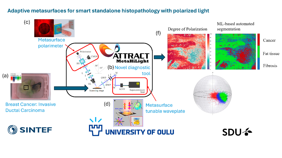
Picture caption*
The MetaHiLight project is focused on developing a novel diagnostic technique for cancer. It utilizes the invisible properties of light (polarization) to provide faster and more detailed information about tissue samples. Leveraging chip-based technology, the developed optical technologies enable miniaturization and large-scale production for low-cost and portable diagnostic devices. The research team has been selected to showcase this technology through demos at the Innovation Village of the SPIE Photonics Europe 2024 conference held in Strasbourg from April 7th to 11th.
This event brings together experts from the entire field of optics and photonics, presenting technologies ranging from digital optics to quantum technologies, optical imaging, sensors, and metrology, from THz photonics and 3D printed optics to quantum optics and biomedical optics, among others. From April 9th to 10th, more than 100 companies will present in the exhibition area and the MetaHiLight team will be showcasing breakthrough technologies for polarimetric imaging of tissue (based on its advances) at Booth 3 in the Innovation Village.
“We are grateful to be part of the SPIE Photonics Europe as it is a premier event, and I believe that participating here is a great opportunity for us as we will be able to show our more recent advances and also, we will be able to connect with people that could be interested in collaborating with the project,” explained Christopher Dirdal, coordinator of the project.
MetaHiLight is an R&D&I project within the ATTRACT ecosystem and is coordinated by SINTEF, one of Europe’s largest research institutes, in partnership with the Oulu University and the University of Southern Denmark.
For additional information about the project, visit here.
* Picture caption:
The MetaHiLight project at a glance. Treatment decisions for many pathological conditions, including inflammatory, degenerative, autoimmune, infectious diseases, and cancer, are largely based on the microscopy study of tissue samples (histology). A tissue sample is shown (a). The MetaHiLight project develops a system (b) which utilizes contrast not available to the human eye (polarization of light) for which it is possible to avoid several time-consuming steps in preparing tissue samples (in the classical histology workflow), thereby significantly speeding up diagnosis. Breakthrough novel metasurface components are developed to ensure this: (c) a metasurface-based polarimeter, and (b) a metasurface-based tunable waveplate. The resulting diagnostic tool provides a polarization map of the tissue samples (f): Whereas the conventional approach for examining tissue samples strongly relies on the skills and qualifications of the pathologist performing the examination, using machine learning (ML) based automated segmentation provides a digital technique for histology, thus avoiding inter-observer variability.”


